While the brain is assumed to exert homeostatic functions to keep the cellular energy status constant under physiological conditions, this has not been proven. Akiyo Natsubori and Makoto Honda in the Sleep Disorders Project of Tokyo Metropolitan Institute of Medical Science, in collaboration with Tomomi Tsunematsu in Tohoku University, discovered a cortex-wide variation of intracellular concentrations of ATP, the major cellular energy metabolite, in excitatory neurons across the sleep-wake states in mice. The ATP levels decreased during non-REM sleep and greatly decreased during REM sleep compared with the wake state, whereas the cerebral hemodynamics for energy supply greatly increased during REM sleep. These suggest a negative energy balance in cortical neurons during REM sleep due to high cellular energy-consuming activities. These indicate that cerebral energy metabolism may not always meet neuronal energy demands.
It is assumed that the brain has homeostatic mechanisms to prevent the depletion of cellular energy, required for all cellular activities. For example, the blood flow increases, and oxygen and glucose are actively delivered in the brain region in which neural firing activity occurs, which mechanisms have been used for functional brain imaging such as fMRI and PET. Besides, the cerebral blood flow and glucose uptake into the cells fluctuate accompanying the variations of cellular activities in the brain across the sleep-wake states. Under these brain energy homeostatic mechanisms, it is hypothesized that the cellular energy status in the brain could be maintained constant in all physiological conditions including across the sleep-wake states of animals. However, this has not been experimentally proven.
The research group conducted in vivo optical recordings of intracellular concentration of adenosine 5’-triphosphate (ATP), the major cellular energy metabolite, using a genetically-encoded fluorescent probe (ATeam: Imamura et al., 2009, PNAS) in the mouse brain. Using a fiber photometry and wide-field microscopy, they demonstrated a cortex-wide variation of cytosolic ATP levels in the cortical excitatory neurons depending on the sleep-wake states of animals: The ATP levels were high during the wake state, decreased during non-REM sleep, and profoundly decreased during REM sleep (Figure 1). On the other hand, cerebral blood flow, as a metabolic parameter for energy supply, slightly increased during non-REM sleep and greatly increased during REM sleep, compared with the wake state. The reduction in neuronal ATP levels was also observed under general anesthesia in mice and response to local brain electrical stimulation for neuronal activation, whereas the hemodynamics was simultaneously enhanced.
Since the neuronal ATP levels increase throughout the cortex during wake state, which is when the cellular energy demand increases, brain mechanisms for energy modulation could increase the neuronal ATP levels in a cortex-wide manner in response to the sleep-to-wake transition of animals. Meanwhile, the great reduction of neuronal ATP levels during REM sleep despite a simultaneous increase of cerebral hemodynamics for energy supply suggests negative energy balance in neurons, which could be due to REM sleep-specific promotion of energy-consuming activities such as heat production (Figure 2). Eventually, cerebral energy metabolism may not always meet neuronal energy demands, consequently resulting in physiological fluctuations of intracellular ATP levels in neurons.
The researches will continue to investigate the neuronal energy-consuming activity as the cause of cellular ATP reduction during REM sleep. The significant reduction of ATP levels in the cortical excitatory neurons during REM sleep is useful as a novel biomarker of REM sleep. Furthermore, it is expected to elucidate brain metabolic functions which bring state-dependent variation of neuronal ATP levels.
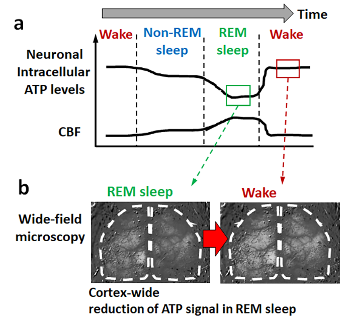
(a) The ATP levels were high during the wake state, decreased during non-REM sleep, and greatly decreased during REM sleep. The CBF showed a variation in the opposite direction to ATP. (b) Wide-field microscopic imaging revealed the cortex-wide variations in neuronal ATP levels across the sleep-wake states.
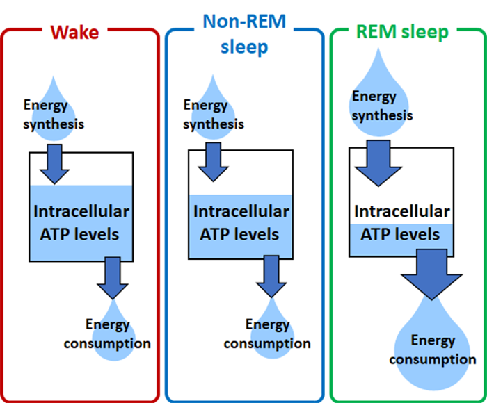
During REM sleep, brain energy synthesis activities (measured as cerebral blood flow) increased while neuronal ATP levels significantly decreased, suggesting a great increase in neuronal energy-consuming activities.