Menu
Tomita Y, Toriumi K, Miyashita M, Suzuki K, Kawaji H, Itokawa M, Arai M. Distinct microRNA profiles in neuron-derived extracellular vesicles between recent-onset and chronic-phase schizophrenia. Schizophrenia (2025) in press.
doi: 10.1038/s41537-025-00706-x
Schizophrenia often emerges from adolescence to early adulthood, a critical neurodevelopmental period during which the brain undergoes rapid maturation. Understanding the molecular changes occurring in the brain at the time of onset is therefore essential for elucidating the pathophysiology of the disorder. However, traditional studies using postmortem brain tissue have been unable to directly capture these early-stage molecular dynamics.
MicroRNAs (miRNAs) play key roles in neurogenesis and synapse formation, and their involvement in schizophrenia has been suggested. Nevertheless, miRNA alterations specifically during the early phase of illness remain poorly understood. Recently, neuron-derived extracellular vesicles (NEVs) isolated from blood have attracted attention as a minimally invasive method for accessing brain-derived molecular information. NEVs contain miRNAs whose profiles are thought to reflect the molecular state of neurons.
In this study, we extracted NEVs from patients with recent-onset schizophrenia (within 5 years of onset) and those in the chronic phase, and compared their miRNA profiles to identify onset-specific molecular alterations and potential mechanisms of disease progression.
(A) PEN was synthesized in vitro by incubating human plasma with GlcA. A total of 93 natural compounds (C1–C93) were screened by adding them to the PEN synthesis system, and the amount of PEN synthesized was expressed as a relative value with the control set to 1. Compounds with a flavonoid backbone exhibited strong inhibitory activity. (B) The chemical structure of Petunidin chloride (C1), the compound with the strongest inhibitory effect, is shown.
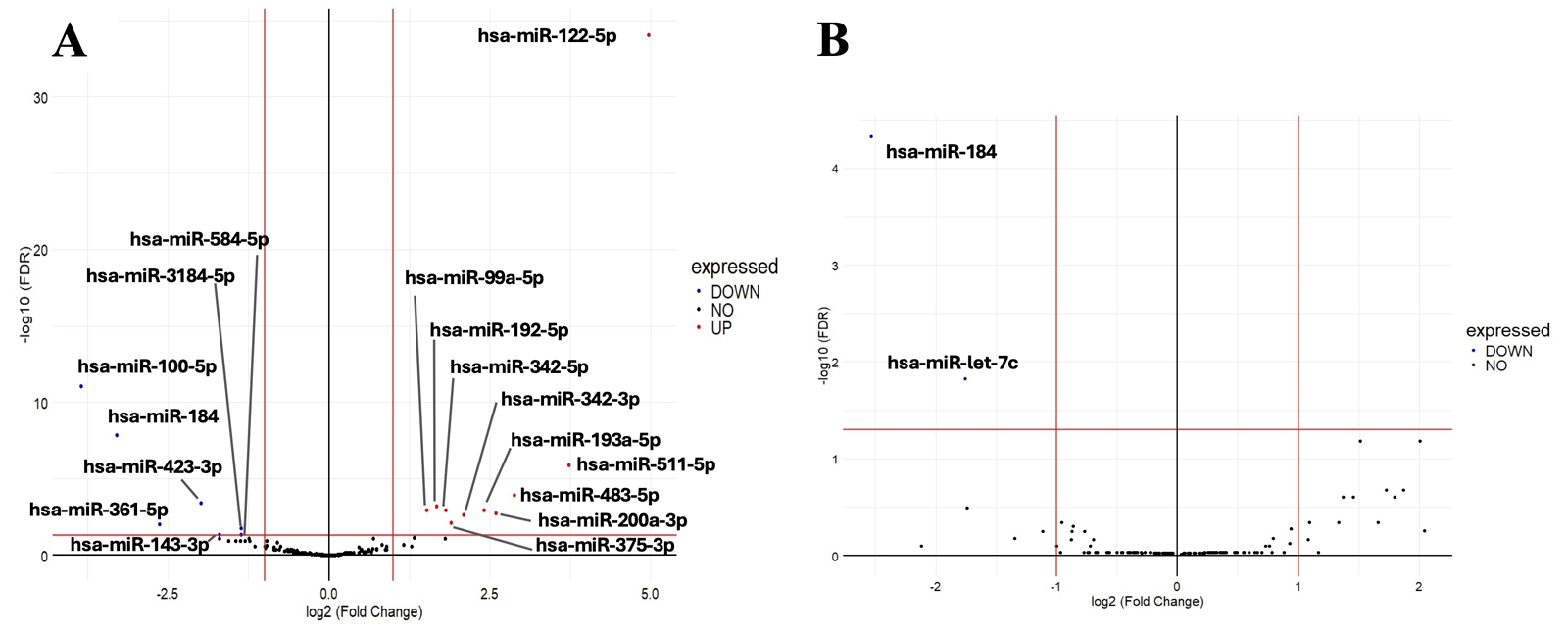
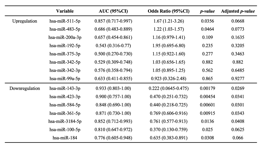
To assess biomarker potential, we conducted univariate logistic regression analyses on 15 of the 17 ROS-associated miRNAs, excluding two that were influenced by medication dosage (Table 1). Among them, miR-143-3p demonstrated the strongest diagnostic performance, with an AUC of 0.933, as well as high sensitivity, specificity, and accuracy. Functional analyses further revealed that target genes of miRNAs altered in the ROS group were enriched for pathways related to neuronal projection development and endosomal transport, whereas miRNAs altered in the CS group were associated with ER–nuclear signaling and regulation of postsynaptic receptor levels. These findings indicate that miRNAs exert distinct functional impacts during the early and chronic stages of schizophrenia.
This study represents the first attempt to characterize miRNA expression in NEVs during the immediate onset of schizophrenia, demonstrating stage-specific molecular mechanisms and highlighting the central role of neurodevelopmental abnormalities in disease onset. Additionally, our results underscore the potential of NEVs as a minimally invasive tool for accessing brain-derived molecular information and suggest that early-altered miRNAs may serve as promising biomarkers for early diagnosis.