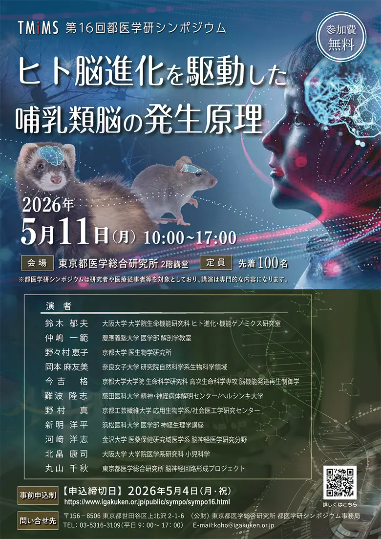
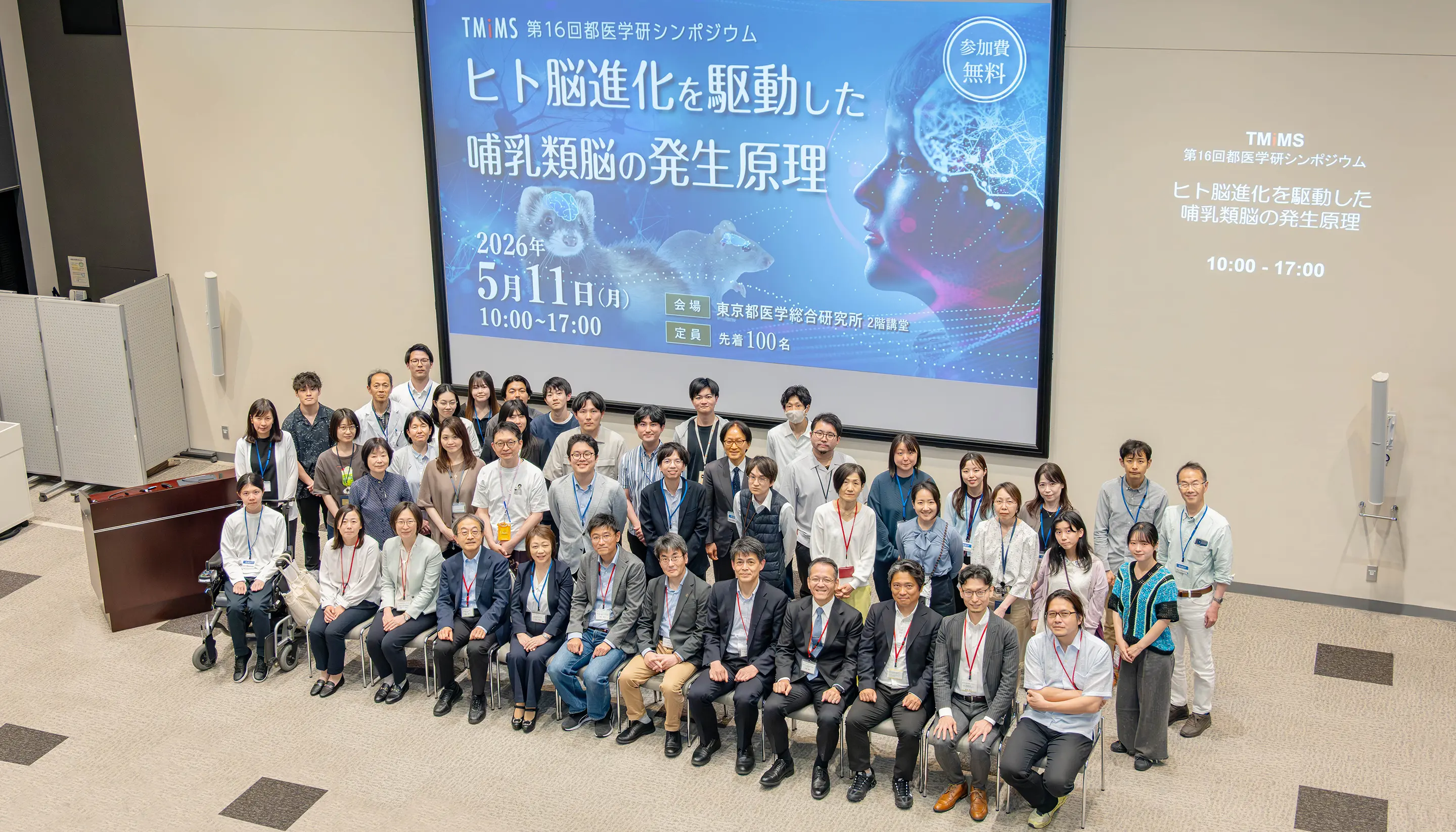
background_sympo20

News

2025

  • July 30-Augst 1, 2025: We held a summer seminar on brain development and evolution for high school students. (Photo)
  • July 24-27, 2025: Dr. Maruyama attended, researcher Hatanaka and trainees Yamazaki and Shinohara gave a poster presentation at the 48th Annual Meeting of the Japan Neuroscience Society. (Photo)
  • July 14-16, 2025: Dr. Maruyama gave an oral presentation as an invited speaker and trainee Achiwa gave a poster presentation at the Anatomical Society Summer Meeting held at St. John's College, University of Oxford, UK. (Photo)
  • June 27-28, 2025: The 4th research group meeting of AMED-CREST “Study on the dynamism of subplate neural activity during brain development” was held at TMIMS. (Photo)
  • June 4-8, 2025: Dr. Maruyama gave an oral presentation as an invited speaker at Black Sea Neurogenesis 2025, Bulgaria. (Photo)
  • June 2-3, 2025: Dr. Maruyama gave a lecture as a Neuroscience Theme Guest Speaker at Sherrington Library, University of Oxford, UK. (Photo)
  • May 25-28, 2025: Dr. Maruyama gave an oral presentation, researchers Moriya and Matsumura, and trainee Sugita gave a poster presentation at Cortical Development Conference 2025, Sicily, Italy. Trainee Sugita received the Best Question Award. (Photo)
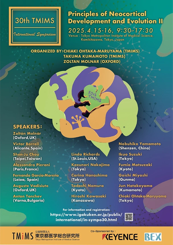
  • April 15-16, 2025: Dr. Maruyama, Dr. Kumamoto and Prof. Molnar organized the 30thTMIM International Symposium “Principles of Neocortical Development and Evolution II” at our institute. (Photo)
  • April, 2025: Dr. Maruyama, Dr. Kumamoto and former researcher Hara at Research Center for Genome & Medical Science published a paper in Scientific Reports on “The spatial transcriptome of the late-stage embryonic and postnatal mouse brain reveals spatiotemporal molecular markers.”
  • April, 2025: Dr. Maruyama and guest researcher Nomura published a paper in Development, Growth & Differentiation on “Genetic and developmental bases for mammalian neocortical evolution.”
  • April 3-5, 2025: Dr. Maruyama and Prof. Molnar gave a lecture at Brain Development Symposium Kyoto 2025, hosted by Kyoto Women’s University. (Photo)
  • March, 2025: Prof. Zoltan Molnar gave a lecture at TMIMS Seminar as follows: (Photo)
    • March 10: Development and Evolution of Thalamocortical Connectivity
    • March 27: Transient Circuits in the Developing Brain
  • March, 2025: Prof. Zoltan Molnar gave anatomy lecture series at TMIMS as follows: (Photo)
    • March 13: Lectures on neuroanatomy-I: Thomas Willis (1621-1675) The Founder of Neuroanatomy and Clinical Neurology
    • March 26: Lectures on neuroanatomy-II: Insight into the Life and Work of Sir Charles Sherrington(1857-1952)
    • April 9: Lectures on neuroanatomy-III: Evolutionary Developmental Biology of the Mammalian Cerebral Cortex
  • March 9-May 7, 2025: Dr. Maruyama invited Prof. Zoltan Molnar from the University of Oxford, as a part of Foreign Researcher Invitation Program. (Photo)
  • March 12, 2025: Trainee Nomura received the Excellent Presenter Award at the 3rd section of the institute's research presentation meeting. (Photo)
  • Feb. 22, 2025: Dr. Maruyama gave a lecture titled “What is the brain and how is it made?” to the general public at the 8th Tomin Koza (public lecture series) on “How the brain was born and evolved - the evolutionary path to the human brain and disease.” (Photo)
  • Jan. 16-17, 2025: Dr. Maruyama gave an oral presentation online, researcher Matsumura gave a poster presentation at AMED Area Conference, Fukuoka.

2024

  • Nov. 27-29, 2024: Dr. Maruyama gave an oral presentation as an invited speaker and trainees Song, Nomura, Ishizuka and Doi gave a poster presentation at the 47th Annual Meeting of the Molecular Biology Society of Japan.
  • Sept. 12, 2024: Dr. Maruyama gave an oral presentation as an invited speaker at Evolution and Development of Nervous System, Croatia.
  • July 31- Augst 2: We held a summer seminar on brain development and evolution for high school students. (Photo)
  • July 24-27, 2024: Dr. Maruyama and trainee Sugita gave an oral presentation and researcher Moriya and trainees Achiwa and Wada gave a poster presentation at the 47th Annual Meeting of the Japan Neuroscience Society.
  • June 23, 2024: Prof. Debra Lynn Silver of Duke University held a seminar at the TMIMS. She gave technical guidance to the researchers. (Photo)
  • June 14-15, 2024: The 3rd research group meeting of AMED-CREST “Study on the dynamism of subplate neural activity during brain development” was held at TMIMS.(Photo
  • June 13, 2024: Dr. Maruyama and former trainee Kaneko published a paper in EMBO reports on “ADAMTS2 promotes radial migration by activating TGF-βsignaling in the developing neocortex.”
  • May 9, 2024: Dr. Maruyama, trainee Katayama and colleagues published a paper in the Journal of Comparative Neurology on “Thalamic activity-dependent specification of sensory input neurons in the developing chick entopallium.”
  • April 19, 2024: Dr. Maruyama received the Prize for Science and Technology (Research Category) by the Minister of Education, Culture, Sports, Science and Technology for 2024. (Photo)
  • March 16, 2024: Trainee Katayama received the Best Presentation Award at the 76th Annual Meeting of the Kanto Branch of the Zoological Society of Japan.(Photo
  • March 11, 2024: Trainee Morioka received the Excellent Presenter Award at the 3rd section of the institute's research presentation meeting.
  • February 17, 2024: Trainee Sugita received the Best Presentation Award at the winter seminar co-hosted by the Kinki and Chugoku-Shikoku Chapters of the Young Researchers Society of Biochemistry.

How are the elaborate neural circuits formed during the development?


This is a profound problem that continues to present new challenges. Neural circuit formation proceeds under diverse cell-cell and cell-extracellular matrix interactions. This project aims to elucidate the molecular mechanisms of such interactions using the mouse cerebral cortex and the Drosophila neuromuscular junction as models.

Cortical Development Research Team

Our lab studies the principles of neocortical development. 「How is the elaborate neural network of the brain organized?」There are more questions than answers. Although organic dysfunctions of neocortical structure cause various neurological and psychiatric disorders, the development mechanism remains unknown. We have been tackling this problem with the standpoint of “understanding the principle of brain construction”. The cerebral neocortex is responsible for higher brain functions, such as conscious thought and language in humans. In the neocortex, neurons are arranged in a precise 6-layered structure formed by the sequential generation of neurons and their accurate migration toward the brain surface during the fetal period. How is this migration controlled? We study subplate neurons, which develop and mature extremely early during cortical development, to answer this question. Subplate neurons play an essential role in establishing neuronal connections between the thalamus and the neocortex. However, we recently found that subplate neurons also play a crucial role in radial neuronal migration by interacting directly with young migrating neurons. Moreover, the subplate layer is surrounded by a rich extracellular matrix (ECM), suggesting a vital signalling center for mammalian corticogenesis. Understanding the various functions of subplate neurons will lead to a better understanding of brain development.


The subplate layer lies beneath the cortical plate and is where subplate neurons, the first neurons to differentiate in the neocortex, are distributed (Fig. 2, Refs. 1, 3 ). Although subplate neurons have been known to play an essential role in thalamocortical projection, we recently found that subplate neurons facilitate mode conversion of neuronal migration through the transient synaptic transmission to migrating neurons (Fig. 4, Ref. 4). To create an elaborate neocortex within the limited developmental time of the fetal period, it is necessary for the neogenesis of neurons, migration, and the formation of neural circuits by axonal projection to proceed simultaneously and in synchrony with each other, but the overall control mechanism has remained unclear. In addition to the new functions found in this study, subplate neurons may perform various brain construction processes and play a commanding role in forming the neocortex.


In addition, the neocortex is a unique region acquired by mammals during the process of brain evolution. The cerebrum of birds and reptiles, which do not have this region, does not have a subplate layer. This suggests that the subplate layer played an essential role in the evolution of the neocortex (Fig. 5). To comprehensively identify the signalling pathways that contribute to neuronal migration, we search for a group of genes selectively expressed in migrating neurons and subplate neurons using DNA microarrays, single-cell RNAseq, and other methods.

We are also interested in abundant proteoglycans and other extracellular matrix components expressed in the subplate layer. Although chondroitin sulfate proteoglycans are expressed in the cerebral cortex, including the subplate layer, during development when neuronal migration is actively ongoing, their functions have not been clarified. We have been focusing on the role of chondroitin sulfate proteoglycans in the formation of the cerebral cortex. Chondroitin sulfate is a linear polysaccharide consisting of a large number of polymerized disaccharide units of glucuronic acid and N-acetylgalactosamine, and the sulfation of the constituent sugars and the C5 epimerization of glucuronic acid gives rise to a great variety of structures in the sugar chain (Fig. 6). Among them, the region with high sulfate density, rich in polysulfated forms, is assumed to contribute to the binding of various proteins and is expected to be functionally important. We have investigated the role of the glycosylation modifying enzymes; GalNAc4S-6ST and C2ST using in utero electroporation. As a result, when these enzymes were knocked down, the migration of neurons was impaired, and they became stagnant in the subventricular zone (Ref. 5). The polysulfated structure of chondroitin sulfate is necessary for the morphological change of neurons from multipolar to bipolar during radial migration. In addition, hippocampal neurons were cultured in chondroitin sulfate-degrading enzyme (chondroitinase ABC), and projection elongation was observed. As a result, degradation and removal of chondroitin sulfate caused abnormalities in nerve polarization, and many neurons grew multiple axons (Ref. 6). Moreover, by observing radial migration in slice cultures, it was found that the leading processes of migrating cells were difficult to determine and that multipolar-bipolarity conversion was impaired (Fig. 7). In the future, we would like to analyze how the process of cortical construction is regulated by proteoglycans and to discuss the role of extracellular matrix regulation in neocortical development and evolution.


References

(1) Ohtaka-Maruyama C and Okado H.(2015). Molecular pathways underlying projection neuronproduction and migration during cerebral cortical development. Front Neurosci. 9:447, doi: 10.3389/fnins.2015.00447. eCollection 2015.

(2) Maeda N. (2015). Proteoglycans and neuronal migration in the cerebral cortex during development and disease.Front. Neurosci. 9:98, doi: 10.3389/fnins.2015.00098.

(3) Ohtaka-Maruyama C., Hirai S., Miwa A., Heng J.I., Shitara H., Ishii R., Taya C., Kawano H., Kasai M., Nakajima K. and Okado H. (2013). RP58 regulates the multipolar-bipolar transition of newborn neurons in the developing cerebral cortex. Cell Rep. 3, 458-471.

(4) Ohtaka-Maruyama C., Okamoto M., Endo K., Oshima M., Kaneko N., Yura K., Okado H., Miyata T., Maeda N.(2018).Synaptic transmission from subplate neurons controls radial migration of neocortical neurons. Science 360, 313-317.

(5) Ishii M., and Maeda N. (2008). Oversulfated chondroitin sulfate plays critical roles in the neuronal migration in the cerebral cortex.J. Biol. Chem. 283, 32610-32620.

(6) Nishimura K., Ishii M., Kuraoka M, Kamimura K. and Maeda N. (2010). Opposing functions of chondroitin sulfate and heparan sulfate during early neuronal polarization. Neuroscience 169, 1535-1547.

People


People

Project leader

chief
Chiaki Ohtaka-Maruyama
Technical staff
Kumiko Hirai
Aiko Odajima
Yoshiko Takahashi
Illia Aota
Guest Researchers
Nobuaki Maeda
( Former project leader)
Kei Yura
(Professor, Ochanomizu Univ)
Tadashi Nomura
(Associate Professor,Kyoto Prefectural University of Medicine)
Carina Hanashima
(Professor, Waseda Univ.)
Yoshiko Honda
(Associate Professor, Tokyo Women’s Medical Univ.)

Publications


  • 2024

    Kaneko N, Hirai K, Oshima M, Yura K, Hattori M, Maeda N, Ohtaka-Maruyama C. ADAMTS2 promotes radial migration by activating TGF-β signaling in the developing neocortex. EMBO reports 2024 Jul;25(7):3090-3115. doi: 10.1038/s44319-024-00174-x. (査読有)

    Katayama R, Kumamoto T, Wada K, Hanashima C, Ohtaka-Maruyama C. Thalamic activity-dependent specification of sensory input neurons in the developing chick entopallium. J Comp Neurol. 2024 Jun;532(6):e25627. doi: 10.1002/cne.25627.(査読有)

  • 2023

    (1) Hara Y†, Kumamoto T†, Yoshizawa-Sugata N, Hirai K, Song X, Kawaji H*, Ohtaka-Maruyama C*. “Spatial transcriptome of developmental mouse brain reveals temporal dynamics of gene expressions and heterogeneity of the claustrum.” bioRxiv (2023) 2023.04.12.536360

    (2) Hirai S, Miwa H, Shimbo H, Nakajima K, Kondo M, Tanaka T, Ohtaka-Maruyama C, Hirai S, Okado H. “The mouse model of intellectual disability by ZBTB18/RP58 haploinsufficiency shows cognitive dysfunction with synaptic impairment.” Mol Psychiatry. (2023) Feb 1.

  • 2022

    (1) Kaneko N, Hirai K, Oshima M, Yura K, Hattori M, Maeda N, Ohtaka-Maruyama C. “ADAMTS2 regulates radial neuronal migration by activating TGF-β signaling at the subplate layer of the developing neocortex.” bioRxiv (2022) 2022.08.07.502954

    (2) Takigawa-Imamura H, Hirano S, Watanabe C, Ohtaka-Maruyama C, Ema M, Mizutani KI. “Computational Model Exploring Characteristic Pattern Regulation in Periventricular Vessels.” Life (Basel). (2022) Dec 9;12(12):2069.

    (3) Tsurugizawa T, Kumamoto T, Yoshioka Y. “Utilization of potato starch suspension for MR-microimaging in ex vivo mouse embryos.” iScience. (2022) Nov 30;25(12):105694.

    (4) Kumamoto T*, Ohtaka-Maruyama C. Visualizing cortical development and evolution: a toolkit update. Front. Neurosci. (2022) Apr 12;16:876406.

  • 2021

    (1) Miyatake S†., Kato M†., Kumamoto T., Hirose T., Koshimizu E., Matsui T., Takeuchi H., Doi H., Hamada K., Nakashima M., Sasaki K., Yamashita A., Takata A., Hamanaka K., Satoh M., Miyama T., Sonoda Y., Sasazuki M., Torisu H., Hara T., Sakai Y., Noguchi Y., Miura M., Nishimura Y., Nakamura K., Asai H., Hinokuma N., Miya F., Tsunoda T., Togawa M., Ikeda Y., Kimura N., Amemiya K., HorinoA., Fukuoka M., Ikeda H., Merhav G., Ekhilevitch N., Miura M., Mizuguchi T., Miyake N., Suzuki A., Ohga S., Saitsu H., Takahashi H., Tanaka F., Ogata K., Ohtaka-Maruyama C., Matsumoto N.(2021). Denovo ATP1A3 variants cause polymicrogyria. Science Advances 7, 2368.

    (2) Kamimura K., Maeda N.(2021). Glypicans and Heparan Sulfate in Synaptic Development, Neural Plasticity, and Neurological Disorders. Front. Neural Circuits 15,595596.

  • 2020

    (1) Nomura T., Ohtaka-Maruyama C., Kiyonari H., Gotoh H., Ono K. (2020). Changes in Wnt-dependent neuronal morphology underlie the anatomical diversification of neocortical homologues in amniotes. Cell Reports, 31,107592.

    (2) Ohtaka-Maruyama C. (2020). Subplate neurons as an organizer of mammalian neocortical development. Front. Neuroanat.14,8.

  • 2019

    (1) Kamimura K., Odajima A., Ikegawa Y., Maru C., Maeda N. (2019). The HSPG Glypican Regulates Experience-Dependent Synaptic and Behavioral Plasticity by Modulating the Non-Canonical BMP Pathway. Cell Reports 28, 3144-3156.

  • 2018

    (1) Ohtaka-Maruyama C., Okamoto M., Endo K., Oshima M., Kaneko N., Yura K., Okado H., Miyata T., Maeda N.(2018).Synaptic transmission from subplate neurons controls radial migration of neocortical neurons. Science 360, 313-317.

  • 2016

    (1) Sakuma C., Saito Y., Umehara T., Kamimura K., Maeda N., Mosca T.J., Miura M., Chihara T. (2016). The Strip-Hippo pathway regulates synaptic terminal formation by modulating actin organization at the drosophila neuromuscular synapses. Cell Rep. 16, 2289-2297.

    (2) Nomura T., Ohtaka-Maruyama C., Yamashita Y., Wakamatsu Y., Murakami Y., Calegari F., Suzuki K., Gotoh H., Ono K. (2016). Evolution of basal progenitors in the developing non-mammalian brains. Development 143, 66-74.

    (3) Ohtaka-Maruyama C., Nakajima K., Pierani A., Maeda N. (2016). Editorial: Mechanisms of neuronal migration during corticogenesis. Front. Neurosci. 10, 172. Doi:10.3389/fnins2016.00172.

  • 2015

    (1) Heng J.I., Qu Z., Ohtaka-Maruyama C., Okado H., Kasai M., Castro D., Guillemot F, Tan S.S. (2015). The zinc finger factor RP58 negatively regulates Rnd2 for the control of neuronal migration during cerebral cortical development. Cerebral Cortex 25, 806-816.

    (2) Maeda N. (2015). Proteoglycans and neuronal migration in the cerebral cortex during development and disease. Front. Neurosci. 9: 98. (doi:10.3389/fnins.2015.00098)

    (3) Yabe T., and Maeda N. (2015). Histochemical analysis of heparan sulfate 3-O-sulfotransferase expression in mouse brain. Methods Mol. Biol. 1229, 377-387.

    (4) 神村圭亮、前田信明 (2015). 「シナプス形成におけるヘパラン硫酸プロテオグリカンの機能 -ショウジョウバエ神経筋接合部を中心に-」生化学87, 467-470.

    (5) Ohtaka-Maruyama C., Okado H (2015). Molecular pathways underlying projection neuron production and migration during cerebral cortical development. Front Neurosci. 9, 447. Doi:10.3389/fnins.2015.00447.

  • 2014

    (1) Morise J., Kizuka Y., Yabuno K., Tonoyama Y., Hashii N., Kawasaki N., Manya H., Miyagoe-Suzuki Y., Endo T., Maeda N., Takematsu H. and Oka S. (2014). Structural and biochemical characterization of O-mannose-linked human kiler-1 glycan expressed on phosphacan in developing mouse brains. Glycobiology 24, 314-324.

    (2) Kamimura K, Maeda N. (2014). Heparan sulfate proteoglycans in Drosophila melanogaster. In “Glycoscience: Biology and Medicine” , Taniguchi N et al.(ed), Springer, pp. 581-587.

  • 2013

    (1) Kamimura K., Ueno K., Nakagawa J., Hamada R., Saitoe M. and Maeda N. (2013). Perlecan regulates bidirectional Wnt signaling at the Drosophila neuromuscular junction. J. Cell Biol. 200, 219-233.

    (2) Ohtaka-Maruyama C., Hirai S., Miwa A., Hengi J.I., Shitara H., Ishii R., Taya C., Kawano H., Kasai M., Nakajima K. and Okado H. (2013). RP58 regulates the multipolar-bipolar transition of newborn neurons in the developing cerebral cortex. Cell Rep. 3, 458-471.

    (3) 神村圭亮、前田信明 (2013). 「ショウジョウバエのシナプスから眺めたヘパラン硫酸プロテオグリカンの機能」 実験医学31, 1488-1493.

  • 2012

    (1) Yasuda T., Saegusa C., Kamakura S., Sumimoto H. and Fukuda M. (2012). Rab27 effector Slp2-a transports the apical signaling molecule podocalyxin to the apical surface of MDCKII cells and regulates claudin-2 expression. Mol. Biol. Cell 23, 3229-3239.

    (2) Hirai S., Miwa A., Ohtaka-Maruyama C., Kasai M., Okabe S., Hata Y. and Okado H. (2012). RP58 controls neuron and astrocyte differentiation by downregulating the expression of Id1-4 genes in the developing cortex. EMBO J. 31, 1190-1202.

    (3) Ohtaka-Maruyama C., Hirai S., Miwa A., Takahashi A. and Okado H. (2012). The 5’-flanking region of the RP58 coding sequence shows prominent promoter activity in multipolar cells in the subventricular zone during corticogenesis. Neuroscience 17, 481-492.

  • 2011年

    (1) Kamimura K., Maeda N., Nakato H. (2011). In vivo manipulation of heparan sulfate structure and its effect on Drosophila development. Glycobiology21, 607-618.

    (2) Maeda N., Ishii M., Nishimura K., and Kamimura K. (2011). Functions of chondroitin sulfate and heparan sulfate in the developing brain. Neurochem. Res.36, 1228-1240.

  • TOP

Address


〒156-8506
2-1-6 Kamikitazawa Setagayaku, Tokyo

Tel +81-3-6834-2367

Tokyo Metroploitan Institute of Medical Science

Developmental Neuroscience Project Chiaki Ohtaka-Maruyama

Your message has been sent. Thank you!

Developmental Neuroscience Project

© Copyright IGAKUKEN[Neural Network], All rights reserved.