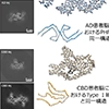
A collaborative research group led by Dr. Masato Hasegawa, Deputy Director and Director of the Department of Clinical Medical Science at the Tokyo Metropolitan Institute of Medical Science, and the MRC Laboratory of Molecular Biology in the UK, has published a paper in Nature demonstrating that "tau filaments from Alzheimer’s disease and corticobasal degeneration convert mouse tau into filaments with the same structures as themselves in the wild-type mouse brain."

A collaborative research group—including Dr. Masato Hasegawa (Deputy Director), Dr. Aki Shimozawa (Chief Researcher, Molecular Pathology and Histology Laboratory), and Dr. Airi Tarutani (Lead Researcher, Dementia Research Project) from the Tokyo Metropolitan Institute of Medical Science, alongside Dr. Sofia Lövestam, Dr. Michel Goedert, and Dr. Sjors H.W. Scheres from the MRC Laboratory of Molecular Biology has demonstrated that injecting tau filaments extracted from the brains of AD or CBD patients into the brains of wild-type mice induces the formation of amyloid-like filaments of mouse tau that share the exact same structure as the injected filaments.
These findings provide concrete evidence that tau, similar to prion strains, propagates and spreads throughout the brain by using a seed (tau filament) as a template. Furthermore, this mouse model has proven to be a highly valuable tool for elucidating the molecular mechanisms by which tau filaments with different structures cause disease-specific pathologies. The results of this study are expected to contribute significantly to the development of novel therapeutic approaches for tauopathies, including AD.
This research article has been published in the journal Nature on September 30 (UK time).
Prions, which are proteinaceous infectious agents, trigger neurodegenerative diseases such as Creutzfeldt-Jakob disease and bovine spongiform encephalopathy. They do this by causing misfolded (abnormal) prion proteins to induce structural changes in normal prion proteins, leading to self-amplification of the abnormal form. Differences in the folding structures of these abnormal prion proteins are believed to give rise to distinct "prion strains," which exhibit different incubation periods and clinical symptoms.
Similarly, tau protein, which is implicated in more than 20 neurodegenerative diseases including AD, is known to form amyloid-like filaments and exhibit prion-like properties in patient brains. To date, cryo-EM structural analyses have been performed on tau filaments extracted from the brains of patients with various tauopathies, revealing that each disease is characterized by a unique folding structure of tau filaments. Furthermore, because identical tau filament structures have been identified across different brain regions within the same patient, it has been suggested that tau filaments with specific structures amplify and propagate within the human brain.
Additionally, "prion-like transmission" has been proposed as the mechanism by which tau lesions spread to widespread brain regions as the disease progresses. Indeed, it has been reported that injecting tau filaments into mouse brains causes tau pathology to spread from the injection site to remote brain regions over time. This phenomenon suggests that tau filaments are released into the extracellular space, taken up by neighboring cells, and transmitted from cell to cell.
However, it had not yet been fully proven whether tau filaments with different structures cause distinct disease pathologies through prion-like mechanisms, or whether different tau strains maintain their structural characteristics during cell-to-cell transmission.
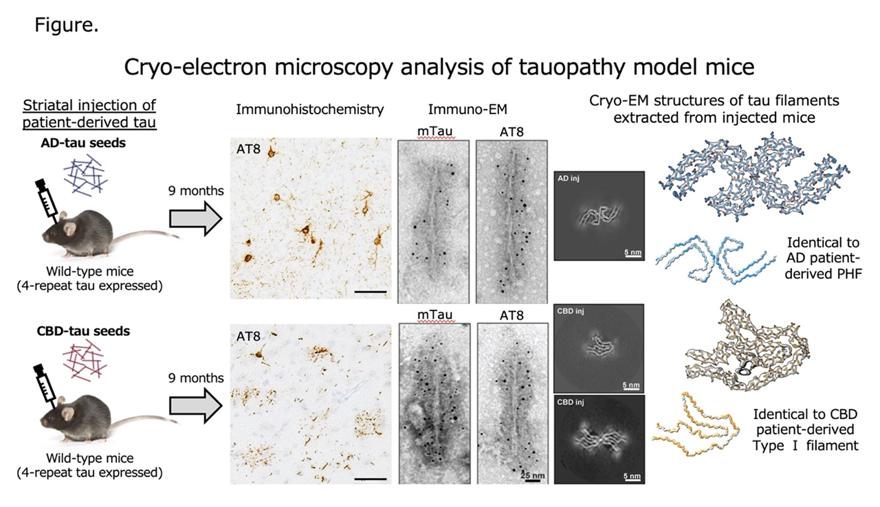
Tau filaments extracted from the brains of AD or CBD patients were injected into the striatum of wild-type mice aged 6 to 18 weeks. Nine months post-injection, immunohistochemical staining using the mTau antibody (which specifically recognizes mouse-derived tau) and the AT8 and AT100 antibodies (which recognize phosphorylated tau) confirmed that tau pathology had spread from the injection site to the cerebral cortex, corpus callosum, and other brain regions. Conversely, these pathologies were not stained by the HT7 antibody, which is specific to human tau.
Furthermore, the differences between the diseases from which the injected tau filaments originated were reflected in the distribution and morphology of the formed pathology. In mice injected with AD-derived tau filaments, tau pathology was observed primarily in the cell bodies and processes of neurons. In contrast, in mice injected with CBD-derived tau filaments, tau pathology was observed not only in neurons but also in glial cells, showing structures resembling astrocytic plaques *6 and coiled bodies *7, which are well-known characteristic pathologies of CBD. These results indicate that structural differences in tau filaments influence not only the morphology of the tau pathology formed in the mouse brain but also the specific cell types affected.
Sarkosyl-insoluble fractions were extracted from the brains of mice injected with patient-derived tau filaments and analyzed by immunoblotting. The results showed that the insoluble tau accumulating in the brain was positive for the mTau antibody but negative for the HT7 antibody. Furthermore, time-course analysis of the injected human tau filaments revealed that they disappeared within approximately one week, while mouse-derived insoluble tau increased in a time-dependent manner. These findings indicate that the tau accumulating in the mouse brain does not consist of the injected filaments themselves, but is derived from endogenous mouse tau.
Further analysis using antibodies that recognize the C-terminal region of tau revealed that the tau formed by injecting AD-derived tau filaments and CBD-derived tau filaments displayed distinct biochemical characteristics. Subsequent immunoelectron microscopy of the insoluble fractions revealed numerous filament structures positive for mTau and AT8, suggesting that the tau filaments accumulating in the mouse brain are indeed composed of mouse tau. Moreover, the brains of mice injected with AD-derived tau filaments formed filaments with the twisted structure characteristic of AD, whereas the brains of mice injected with CBD-derived tau filaments exhibited filament structures with a longer twist.
Cryo-EM analysis of the tau filaments extracted from the mouse brains revealed that the accumulating tau filaments adopted the exact same structures as the injected human tau filaments. In mice injected with AD-derived tau filaments, the majority of the amplified filaments were helical structures consisting of two twisted protofilaments. At a resolution of 3.6 Å, these were shown to be identical in structure to the filaments derived from AD patient brains.
Two types of filaments were identified in the brains of mice injected with CBD-derived tau filaments: approximately 70% were single protofilaments, and the remaining 30% were doublets consisting of two twisted protofilaments. At a resolution of 3.4 Å, the single protofilaments were shown to have the identical structure as Type 1 filaments found in CBD patient brains.
The patient brain tissue samples used in this study were generously provided by Dr. Kazuko Hasegawa (National Hospital Organization Sagamihara National Hospital), Dr. Yuko Saito and Dr. Shigeo Murayama (Tokyo Metropolitan Institute for Geriatrics and Gerontology), Dr. Mari Yoshida (Aichi Medical University), Dr. Hisaomi Suzuki and Dr. Mitsumoto Onaya (National Hospital Organization Shimofusa Psychiatric Medical Center), and Dr. Andrew Robinson (University of Manchester).
All animal experiments in this study were approved by the Animal Care and Use Committee of the Tokyo Metropolitan Institute of Medical Science (Approval No. JP17pc0101006) and conducted in strict accordance with relevant laws, regulations, and guidelines.
This study was supported by subsidies from the Tokyo Metropolitan Government, as well as grants from the Japan Agency for Medical Research and Development (AMED), the Japan Science and Technology Agency (JST), and the Japan Society for the Promotion of Science (JSPS) KAKENHI, among others.
