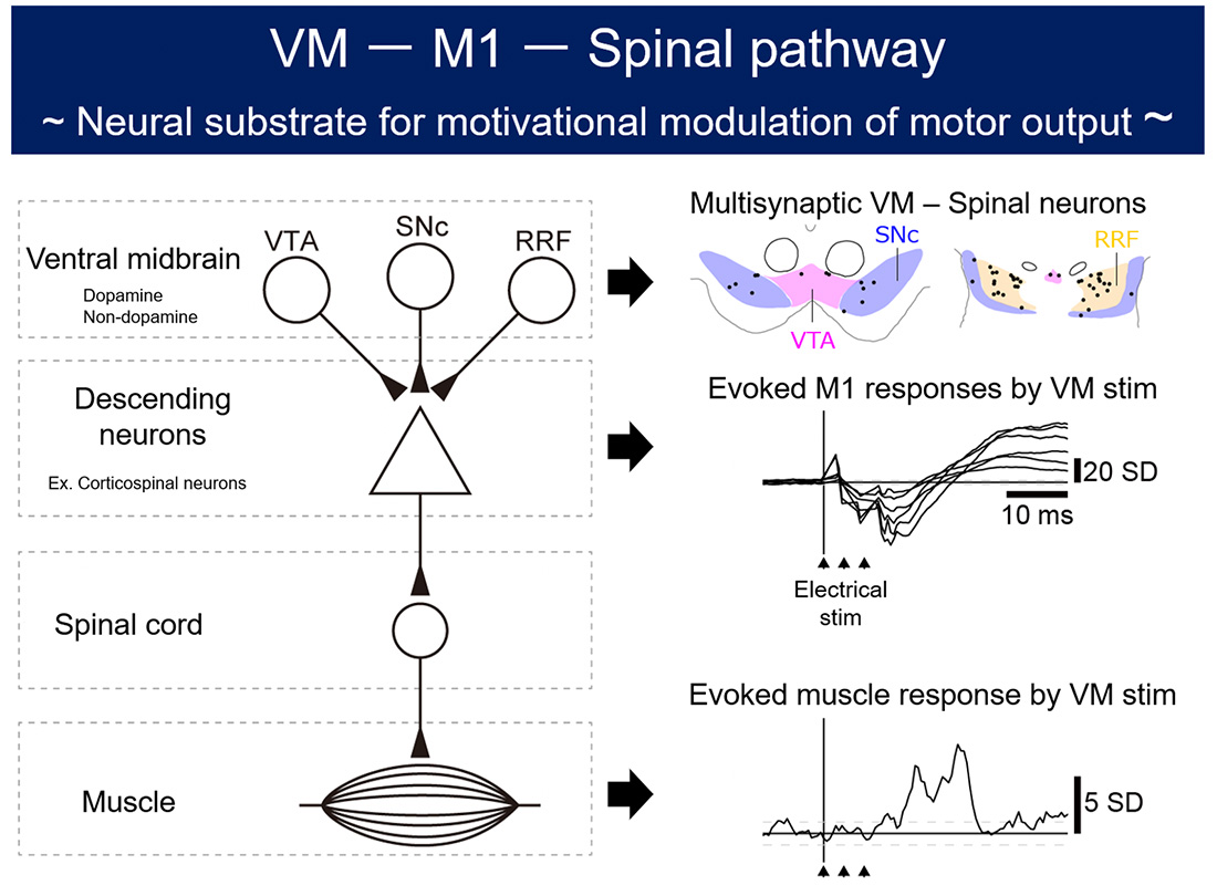
Motivation boosts motor performance. The activity of the ventral midbrain (VM), consisting of the ventral tegmental area (VTA), the substantia nigra pars compacta (SNc), and the retrorubral field (RRF), plays an important role in processing motivation. However, little is known about a neural substrate bridging the VM and the spinal motor output. Here, the research group hypothesized that the VM might exert a modulatory influence over the descending motor pathways. Employing retrograde transneuronal labeling with rabies virus, the research group demonstrated the existence of multisynaptic projections from the VM to the cervical enlargement in an animal model. To investigate the functional significance of the anatomically-identified multisynaptic VM–spinal projections, electrical stimulation was delivered to the VM. Results showed that the VM induced muscle responses in the contralateral forelimb with a few milliseconds delay following the responses of the ipsilateral primary motor cortex (M1). The magnitude of evoked muscle responses was associated with stimulus intensity and pulse number. The muscle responses were diminished during M1 inactivation. Their findings suggest that a multisynaptic VM–M1–spinal pathway may play a pivotal role in modulatory control of the spinal motor output.
This study was supported by the Japan Society for the Promotion of Science (KAKENHI), the Ministry of Education, Culture, Sports, Science and Technology (MEXT) of Japan (a Grant-in-Aid for Scientific Research on Innovative Areas “Brain Information Dynamics”, “Hyper adaptability” and “Adaptive Circuit Shift”:), the Japan Science and Technology Agency (Moonshot R&D), the Cooperative Research Program of Primate Research Institute, Kyoto University and the Cooperative Study Program of National Institute for Physiological Sciences.