
The Intracellular Quality Control Project at the Tokyo Metropolitan Institute of Medical Science has identified a molecular mechanism that helps initiate the formation of autophagic membranes around damaged mitochondria. The study reveals that the small GTPases RAB1A and RAB1B act as molecular links between OPTN, an autophagy adaptor, and ATG9A vesicles, which contribute to the initial formation of autophagic membranes during mitophagy (mitochondria selective autophagy). By recruiting ATG9A vesicles to damaged mitochondria, the OPTN–RAB1 pathway helps establish the site where the autophagic membrane begins to form. The findings were published online in the international scientific journal Autophagy on September 1, 2026.
Mitochondria are essential organelles that produce energy and perform numerous functions required for cell survival. However, damaged mitochondria can become harmful to cells and therefore need to be selectively eliminated. Cells accomplish this through mitophagy, a selective autophagy that recognizes and removes damaged mitochondria. One of the best-characterized mitophagy pathways is controlled by the kinase PINK1 and the E3 ubiquitin ligase Parkin. When mitochondria are damaged, PINK1 and Parkin promote the accumulation of ubiquitin on the mitochondrial surface. This ubiquitin signal is recognized by autophagy adaptors such as OPTN (optineurin), which connect damaged mitochondria to the autophagy machinery. However, an important question has remained: How does an autophagy adaptor such as OPTN initiate the formation of an autophagic membrane around the damaged mitochondria?
The research group previously found that OPTN recruits ATG9A vesicles to damaged mitochondria. ATG9A vesicles are thought to provide membrane seeds and function as important components in the early stages of autophagosome formation. This finding suggested that autophagy adaptors may have a role beyond simply connecting damaged mitochondria to the autophagic machinery. They may also actively recruit the membrane components required to initiate autophagic membrane formation. The molecular mechanism responsible for this recruitment, however, remained unclear.
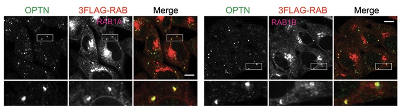
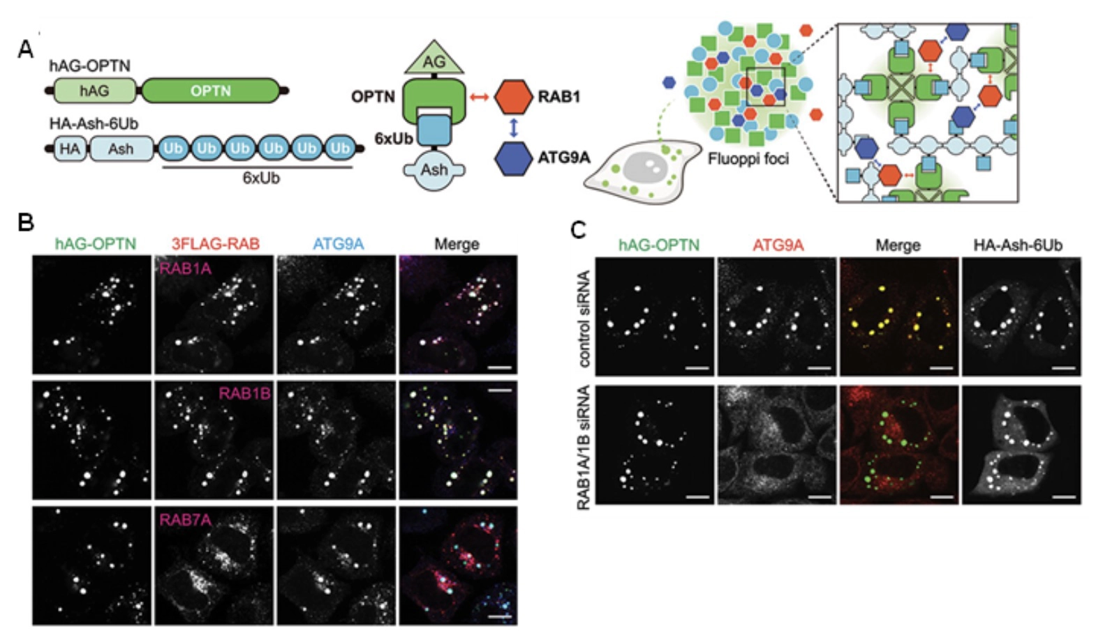
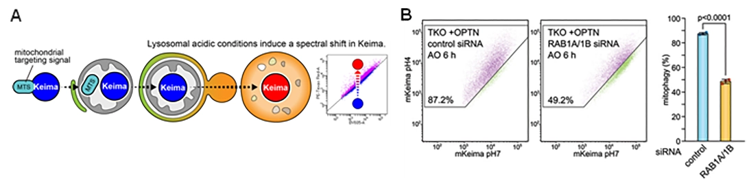
In the present study, the researchers investigated how OPTN recruits ATG9A vesicles during PINK1-Parkin-mediated mitophagy. Humans have approximately 60 RAB GTPases, which function as molecular switches that regulate intracellular membrane trafficking. The researchers screened RAB proteins for interactions with OPTN and identified RAB1A and RAB1B as binding partners of OPTN. The researchers then examined whether RAB1 was required for the recruitment of ATG9A vesicles to damaged mitochondria. When RAB1A and RAB1B were simultaneously knocked down using RNA interference, ATG9A vesicles were no longer efficiently recruited to OPTN sites. Consistent with this defect, suppression of RAB1 reduced PINK1-Parkin-mediated mitophagy. These results indicated that RAB1 is not simply associated with OPTN during mitophagy, but plays an essential role in connecting OPTN to the membrane trafficking machinery required for autophagic membrane formation.
OPTN contains several functional domains that allow it to interact with different components of the mitophagy machinery. The UBAN domain recognizes ubiquitin chains, while other regions interact with autophagy-related proteins. Using an intracellular protein–protein interaction analysis system, structural prediction with AlphaFold, and mutational analyses, the researchers found that OPTN directly interacts with RAB1 through its leucine zipper (LZ) domain. The researchers further found that RAB1 associates with ATG9A vesicles through their C-terminal prenylation. This provides a molecular explanation for how RAB1 can function as a bridge between OPTN on damaged mitochondria and ATG9A vesicles. In this model, OPTN recognizes ubiquitinated damaged mitochondria and simultaneously binds RAB1. RAB1, in turn, associates with ATG9A vesicles, thereby bringing these vesicles into close proximity to the damaged mitochondria. Thus, the study identifies an OPTN–RAB1–ATG9A axis that spatially connects damaged mitochondria with the membrane components required for autophagy initiation.
The findings provide a more complete picture of how mitophagy is initiated. Rather than functioning only as a molecular bridge between ubiquitinated mitochondria and the autophagic membrane, OPTN can also help establish a local membrane-forming site by recruiting ATG9A vesicles through RAB1. This mechanism may be particularly important during the early stages of mitophagy, when the cell must assemble the machinery required to generate an autophagic membrane at the correct location.
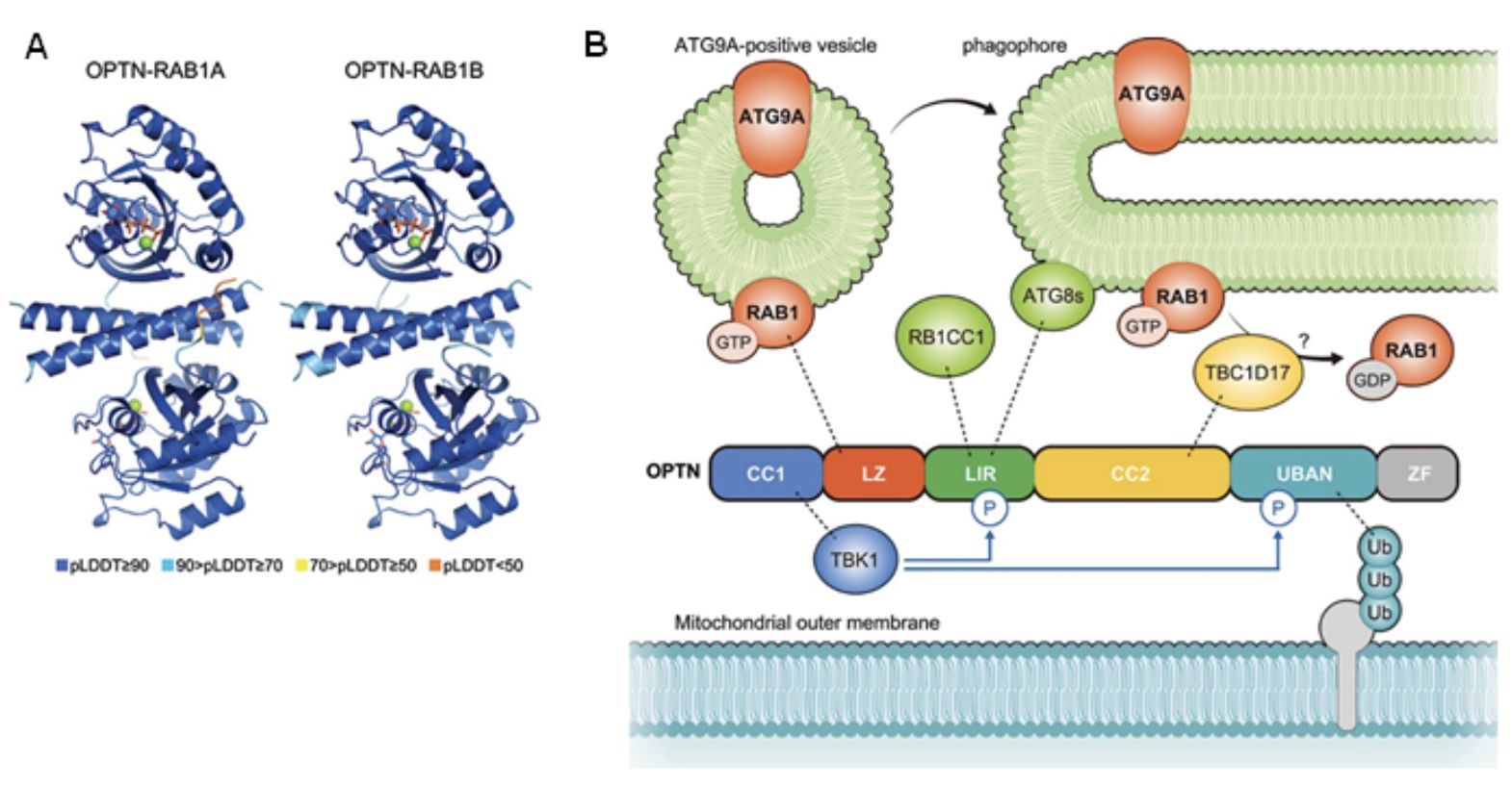
Autophagy is essential for maintaining cellular homeostasis, and defects in autophagy and mitochondrial quality control have been associated with human diseases. Impaired mitophagy is associated with neurodegenerative diseases including Parkinson's disease. PINK1 and Parkin, key regulators of mitophagy, are causal gene products of familial forms of Parkinson's disease. The present study identifies a molecular mechanism that explains how an autophagy adaptor can spatially coordinate mitochondrial ubiquitination with membrane trafficking. By identifying RAB1A and RAB1B as molecular links between OPTN and ATG9A vesicles, the study provides new insight into how the autophagic membrane is initiated at the surface of damaged mitochondria. A better understanding of the fundamental mechanisms controlling mitochondrial quality and autophagy may ultimately contribute to a deeper understanding of diseases associated with defects in these cellular quality-control systems.
This study was supported by the Japan Society for the Promotion of Science (JSPS) KAKENHI (Grant Numbers 26K18571, 19K16068, 21K15084, 24H01894, 18H05501, 23K23841, and 23H04923); the Japan Science and Technology Agency (JST) SPRING (Grant Number JPMJSP2180); the Japan Agency for Medical Research and Development (AMED) CREST (Grant Number JP20gm1410004) and AMED-PRIME (Grant Number JP25gm6910030); ISHIZUE 2023 of Kyoto University; the Grant for the Promotion of Research Activities of The Kyoto University Foundation; the Takeda Science Foundation; Nanken-Kyoten, Medical Research Center Initiative for High Depth Omics, and Multilayered Stress Diseases (Grant Number JPMXP1323015483); the Astellas Foundation for Research on Metabolic Disorders; and the Cell Science Research Foundation.
