TOPICS 2019
-
Yang-Chi-Chun and Hisao Masai (Genome Dynamics Project) published a paper entitled ”Cdc7 activates replication checkpoint by phosphorylating the Chk1 binding domain of Claspin in human cells.” in eLife

-
Hikaru Tsuchiya (Laboratory of Protein Metabolism) published a paper entitled “Structural insights into ubiquitin recognition and Ufd1 interaction of Npl4” in Nature Communications.
-
Miharu Nakanishi (Mental Health Promotion Project) published a paper entitled ”Midlife Psychological Well-Being and its Impact on Cognitive Functioning Later in Life: An Observational Study Using a Female British Cohort.” in Journal of Alzheimer’s Disease
-
Dr. Masato Hosokawa (Dementia Research Project) was awarded the Encouragement Award (Best Basic Research) at the Japan Society for Dementia Research Annual Meeting in 2019.
-
Tetsuya Hirabayashi (Laboratory of Biomembrane)published a paper entitled "SDR9C7 catalyzes critical dehydrogenation of acylceramides for skin barrier formation" in The Journal of Clinical Investigation
-
Yukio Nishimura (Neural Prosthesis Project) published a paper entitled ”Bypassing stroke-damaged neural pathways via a neural interface induces targeted cortical adaptation” in Nature Communications.
-
Takahiko Noro (Visual Research Project) published a paper on glaucoma-like degeneration of the visual system in aged marmosets in Scientific Reports.
-
Dr. Fumika Koyano (Ubiquitin Project) published a paper on “Parkin‐mediated ubiquitylation redistributes MITOL/March5 from mitochondria to peroxisomes” in EMBO Reports.
-
Keisuke Kamimura (Neural Network Project) published a paper entitled ”The HSPG Glypican Regulates Experience-Dependent Synaptic and Behavioral Plasticity by Modulating the Non-Canonical BMP Pathway” in Cell Reports
-
Takahiro Sanada (Viral Infectious Diseases Project) published a paper on “Construction of complete Tupaia belangeri transcriptome database by whole-genome and comprehensive RNA sequencing” in Scientific Reports
-
Yuki Nakayama (ALS Nursing Care Project) published a paper on “Body weight variation predicts disease progression after invasive ventilation in amyotrophic lateral sclerosis” in Scientific Reports
-
Kazuhiko Namekata (Visual Research Project) published a paper on the role of DOCK8 during neurodegeneration in Journal of Biological Chemistry.
-
Daisuke Yamane (Viral Infectious Diseases Project) published a paper on “Basal Expression of Interferon Regulatory Factor 1 Drives Intrinsic Hepatocyte Resistance to Multiple RNA Viruses” in Nature Microbiology
HOME > Topics2019 > 23 July 2019

23 July 2019
Kazuhiko Namekata (Visual Research Project) published a paper on the role of DOCK8 during neurodegeneration in Journal of Biological Chemistry.
DOCK8 is expressed in microglia, and it regulates microglial activity during neurodegeneration in murine disease models.
Summary
DOCK8 is a guanine nucleotide exchange factor whose loss of function results in immunodeficiency, but its role in the central nervous system (CNS) has been unclear. Kazuhiko Namekata and his colleagues in Visual Research Project and Brain Pathology Center in Tokyo Metropolitan Institute of Medical Science demonstrated that DOCK8 is expressed in microglia and regulates microglial activity in disease models of multiple sclerosis (MS) and glaucoma.
- <Title of the paper>
- DOCK8 is expressed in microglia, and it regulates microglial activity during neurodegeneration in murine disease models.
- <Journal>
- Journal of Biological Chemistry
http://www.jbc.org/cgi/doi/10.1074/jbc.RA119.007645
DOI: 10.1074/jbc.RA119.007645
Details
The research group examined the effects of DOCK8 by generating DOCK8 knockout (DOCK8-/-) mice. When experimental autoimmune encephalomyelitis (EAE), a mouse model of MS, was induced in DOCK8-/- mice, clinical symptoms and inflammation in the spinal cords and optic nerves were mild compared with wild-type mice (Figure 1). They found that DOCK8 was expressed in cultured microglia, but not in neurons and glial cells, and that DOCK8 stimulates microglial migration in vitro (Figure 2). These results suggested that inhibition of microglial migration resulted in the reduced severity of EAE. In postmortem brain tissues from MS patients, many DOCK8-positive microglia were detected in the lesion site (Figure 3), implying that DOCK8 may play a role in pathogenesis of MS. They also found that microglial phagocytosis in the retina is impaired in DOCK8-/- mice in the optic nerve injury model, a mouse model of glaucoma (Figure 4). In summary, they have demonstrated that DOCK8 is expressed in microglia and it regulates microglial activity during neurodegeneration. These findings contribute to a better understanding of the molecular pathways involved in microglial activation, and implicate a role of DOCK8 in several neurological diseases.
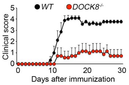
Figure 1 Reduced clinical scores in DOCK8-/- EAE mice.
The severity of EAE, as indicated by clinical scores, is significantly decreased in DOCK8-/- EAE mice (red) compared with WT EAE mice (black).
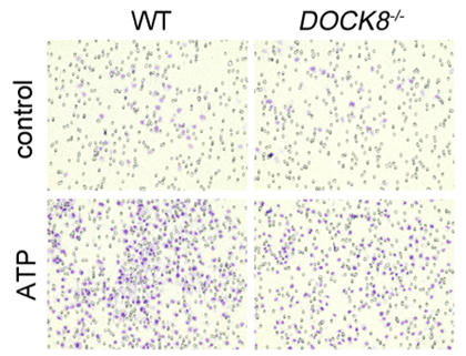
Figure 2 Migration ability of DOCK8-/- microglia is impaired.
ATP-dependent migration ability was examined by counting the number of cells (microglia; purple spots) that have migrated through the membrane using a Boyden chamber assay. Migration ability of DOCK8-/- microglia (bottom right) was significantly lower than WT microglia (bottom left).
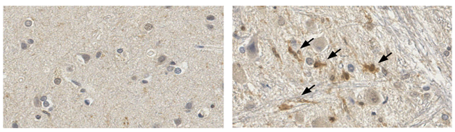
Figure 3 DOCK8 positive microglia are detected in MS patient brain.
Many DOCK8-positive microglia are detected in the lesion site (arrows; right) of the MS patient brain, but not in the non-lesion site (left).
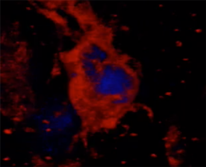
Figure 4 Detection of phagocytic microglia following optic nerve injury.
A retinal ganglion cell (blue) is wrapped and phagocytosed by an activated microglia (red), 5 days after optic nerve injury.